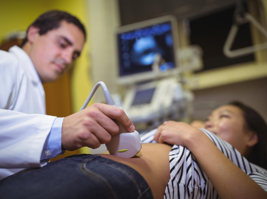
The thyroid gland sits at the front of your neck, just below the Adam’s apple, and it plays a role in almost every function in your body. It regulates metabolism, heart rate, body temperature, energy levels, and much more. When something goes wrong with the thyroid, the effects ripple outward into every system it influences. A thyroid ultrasound is one of the most direct and accurate ways to assess what the gland looks like and whether anything needs attention. At Jhansi Diagnostics & Fetal Medicine Centre, Dr Furquan Ahmad, MBBS MD, performs thyroid ultrasounds as part of a thorough diagnostic process that gives patients and their doctors the clearest possible picture of thyroid health.
Here is when your doctor is likely to order a thyroid ultrasound and exactly what the scan shows.
What Is a Thyroid Ultrasound?
A thyroid ultrasound is a non-invasive imaging scan that uses high-frequency sound waves to produce detailed real-time images of the thyroid gland. No radiation is involved, no injections are required, and the procedure is completely painless. The transducer probe is placed gently on the front of the neck and moved across the surface of the skin to capture images from multiple angles.
The scan takes between 15 and 30 minutes depending on the findings and whether any lymph nodes in the surrounding area need to be assessed alongside the thyroid. Results are available the same day at Jhansi Diagnostics & Fetal Medicine Centre, and Dr Furquan Ahmad explains the findings directly to patients before they leave. You can find out more about the full range of ultrasound scans available at the centre on the dedicated page.
When Does Your Doctor Order a Thyroid Ultrasound?
There are several situations in which a thyroid ultrasound is the appropriate next step. Understanding which category applies to you helps clarify why the scan has been recommended.
The most common reason is the detection of a thyroid nodule. A nodule is a lump or growth within the thyroid gland. Your doctor may feel one during a routine examination of your neck, or it may have been picked up incidentally on another scan such as a neck CT or an MRI done for a different reason. Thyroid nodules are extremely common, affecting a significant proportion of the adult population, and the vast majority are completely benign. However, a small percentage can be malignant, which is why every nodule needs to be assessed properly. A thyroid ultrasound is the first and most important step in that assessment.
A thyroid ultrasound is also ordered when blood tests reveal abnormal thyroid hormone levels. Hypothyroidism, where the thyroid is underactive, and hyperthyroidism, where it is overactive, can both have structural causes that are visible on ultrasound. An enlarged gland, a goitre, or the characteristic appearance of autoimmune thyroid conditions like Hashimoto’s thyroiditis or Graves’ disease can all be identified through imaging.
Symptoms that prompt a thyroid ultrasound referral include a visible or palpable lump in the neck, difficulty swallowing, a persistent hoarse voice, a sensation of pressure or tightness in the throat, and unexplained weight changes combined with other thyroid symptoms. Some patients are also referred for a thyroid ultrasound as part of a routine health check-up, particularly if they have a family history of thyroid disease or thyroid cancer.
Women during or after pregnancy are another group frequently referred for thyroid assessment, as pregnancy places significant demands on thyroid function and can unmask conditions that were previously subclinical. The our services page at Jhansi Diagnostics & Fetal Medicine Centre outlines the full range of diagnostic services available for patients across Jhansi and Bundelkhand.
What Does a Thyroid Ultrasound Actually Show?
A thyroid ultrasound provides detailed information about the gland across several parameters. Understanding what Dr Furquan Ahmad is assessing during the scan helps patients interpret their reports more meaningfully.
The first thing assessed is the size of the thyroid gland. An enlarged thyroid, known as a goitre, can be caused by iodine deficiency, autoimmune conditions, or the presence of multiple nodules. A significantly reduced or atrophied gland can indicate long-standing hypothyroidism or end-stage autoimmune thyroid disease.
The texture of the gland is the next key parameter. A normal thyroid has a uniform, medium-grey echogenicity on ultrasound. In Hashimoto’s thyroiditis, the texture becomes heterogeneous and the overall echogenicity decreases, giving the gland a coarser, patchier appearance. In Graves’ disease, the gland is typically enlarged with increased vascularity, which is visible on Colour Doppler. These textural changes are highly characteristic and allow Dr Furquan Ahmad to identify autoimmune thyroid conditions with considerable accuracy.
Thyroid nodules receive the most detailed assessment during the scan. Each nodule is characterised by its size, shape, margins, composition, echogenicity, and the presence of calcifications or vascularity. These features are used to classify the nodule according to standardised reporting systems that estimate the likelihood of malignancy and guide decisions about whether a biopsy is needed.
Nodules that are purely fluid-filled cysts are almost always benign. Solid nodules with irregular margins, microcalcifications, a taller-than-wide shape, or significant internal vascularity are more suspicious and typically warrant a fine needle aspiration biopsy for cytological analysis. Nodules that fall in between these extremes are monitored with follow-up scans at regular intervals to check for any change in size or characteristics.
The lymph nodes in the neck are also assessed as part of a thorough thyroid ultrasound. Enlarged or abnormal-looking lymph nodes near the thyroid can indicate that a malignant process has spread beyond the gland, which significantly affects staging and management decisions. If you have questions about what the scan involves or how to prepare, the FAQs page covers common queries in detail.
What Happens After a Thyroid Ultrasound?
The next steps after a thyroid ultrasound depend entirely on what the scan finds. If the gland is normal in size, texture, and appearance with no nodules, no further imaging is typically needed unless symptoms persist or blood tests remain abnormal.
If a benign-appearing nodule is found, a follow-up scan is usually recommended after six to twelve months to confirm it is stable in size and appearance. Most benign nodules never cause problems and never require treatment beyond monitoring.
If a nodule has suspicious features, Dr Furquan Ahmad will document these clearly in the report and recommend a fine needle aspiration biopsy. This is a minimally invasive procedure where a thin needle is used to collect a small sample of cells from the nodule for laboratory analysis. The biopsy result determines whether surgical removal is needed or whether continued monitoring is appropriate.
If the scan suggests an autoimmune condition like Hashimoto’s or Graves’ disease, the findings are correlated with the patient’s blood test results and referred to an endocrinologist for medical management. The ultrasound findings in these cases help confirm the diagnosis and establish a baseline for monitoring the gland over time.
Patients at Jhansi Diagnostics & Fetal Medicine Centre receive same-day reports with clear documentation of all findings and recommended next steps. Dr Furquan Ahmad ensures that every patient understands what was found before leaving the centre so that follow-up care can begin without delay. You can book your thyroid ultrasound through the booking appointment page or reach the team through the contact us page with any questions beforehand.
Your thyroid works quietly in the background every day. A single scan is all it takes to know whether it needs attention.