If your doctor has recommended breast imaging and mentioned both a breast ultrasound and a mammogram, you may be wondering what the difference is and why one is being suggested over the other. It’s a question Dr Furquan Ahmad, MBBS MD, hears regularly at Jhansi Diagnostics & Fetal Medicine Centre. The honest answer is that both tests serve different purposes, work better for different types of breast tissue, and in many cases complement each other rather than compete. Understanding what each one does and when each one is appropriate helps you make sense of your doctor’s recommendation and feel confident about the imaging you are getting.
Here is a clear, straightforward comparison of breast ultrasound and mammogram, and guidance on which one is right for your situation.
What Is a Breast Ultrasound?
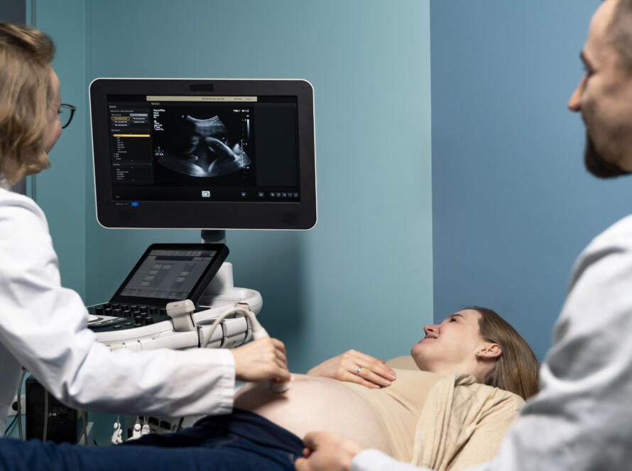
A breast ultrasound uses high-frequency sound waves to produce real-time images of the breast tissue. A handheld probe is moved across the surface of the breast after a small amount of gel is applied to the skin. The sound waves penetrate the tissue and bounce back differently depending on whether they encounter solid tissue, fluid, or fat. The machine converts these returning signals into images that Dr Furquan Ahmad reads to assess the nature and characteristics of any area of concern.
Breast ultrasound involves no radiation whatsoever. It is completely painless, requires no compression of the breast, and produces no discomfort during the procedure. It is particularly well suited to examining specific areas of concern, such as a lump that has been felt during a self-examination or identified on a mammogram, and to distinguishing between solid masses and fluid-filled cysts. You can find out more about the ultrasound services available at Jhansi Diagnostics & Fetal Medicine Centre on the dedicated page.
What Is a Mammogram?
A mammogram is an X-ray of the breast. The breast is placed between two plates that apply firm compression to flatten the tissue, and a low-dose X-ray image is taken. The compression, while uncomfortable for many women, is necessary to spread the breast tissue evenly and produce a clear image. A standard mammogram takes two views of each breast, one from above and one from the side.
Mammograms use a small amount of radiation, although the dose is low and considered safe for routine screening in the appropriate age group. The key strength of mammography is its ability to detect microcalcifications, tiny calcium deposits in the breast tissue that can be an early sign of ductal carcinoma in situ, a very early form of breast cancer, before any lump is palpable or visible on ultrasound. This makes mammography the preferred screening tool for breast cancer detection in women above a certain age with average risk.
The Key Difference: Dense Breast Tissue
This is the most important factor in deciding which test is appropriate, and it is one that is frequently not explained clearly to patients. Breast tissue composition varies significantly between women and changes with age. Younger women typically have denser breast tissue, meaning a higher proportion of glandular and fibrous tissue relative to fat. Older women, particularly post-menopausal women, typically have less dense, fattier breast tissue.
This matters enormously for imaging because dense breast tissue appears white on a mammogram, and so does cancer. When both the tissue and any potential abnormality appear in the same white tone, a mammogram can miss significant findings. This is called the masking effect, and it is the primary reason mammograms are less reliable in younger women and women with dense breasts.
Ultrasound, on the other hand, is not affected by breast density in the same way. It can clearly distinguish a solid mass from surrounding dense tissue regardless of the tissue’s density. This is why Dr Furquan Ahmad at Jhansi Diagnostics & Fetal Medicine Centre recommends breast ultrasound as the primary imaging tool for younger women and for women with known dense breast tissue, while mammography remains the preferred screening tool for older women with less dense breasts.
When Is a Breast Ultrasound the Right Choice?
A breast ultrasound is the appropriate first-line investigation in several specific situations.
For women under 40, breast ultrasound is generally preferred over mammography. Dense breast tissue in this age group makes mammograms less reliable, and the radiation exposure from repeated mammograms over a lifetime is a consideration worth avoiding when ultrasound provides better information anyway.
When a specific lump or area of thickening has been felt by the patient or identified by a doctor during a clinical examination, ultrasound is the most direct way to characterise it. Ultrasound can determine within a single scan whether a palpable lump is a simple benign cyst, which requires no further action, or a solid mass that needs further investigation.
Breast ultrasound is also the appropriate investigation during pregnancy and breastfeeding, as it involves no radiation and does not require breast compression. Women with breast implants benefit from ultrasound imaging as implants can obscure breast tissue on a mammogram.
Any woman with nipple discharge, localised breast pain, skin changes, or axillary lymph node swelling should have a breast ultrasound as part of their initial assessment. The FAQs page at Jhansi Diagnostics & Fetal Medicine Centre has more information about what to expect during a breast ultrasound.
When Is a Mammogram the Right Choice?
Mammography is the established standard for breast cancer screening in women above 40 with average risk and no specific symptoms. Its strength lies in detecting microcalcifications and subtle architectural distortions in fatty breast tissue that ultrasound may not pick up. For post-menopausal women whose breast tissue has become less dense over time, mammography provides a reliable screening picture that is difficult to replicate with ultrasound alone.
Women with a strong family history of breast cancer, particularly those with a first-degree relative diagnosed before 50, are typically advised to begin mammographic screening earlier than the standard recommendation. Women who have previously been treated for breast cancer also require regular mammographic follow-up as part of their long-term monitoring plan.
It is important to note that mammography is a screening tool. It is designed to detect potential problems in women without symptoms. When symptoms are already present, such as a palpable lump, a breast ultrasound is usually the more appropriate starting point regardless of age, often followed by mammography if the ultrasound findings warrant it.
Can Both Tests Be Used Together?
Yes, and in many cases they should be. Breast ultrasound and mammography are complementary investigations, not alternatives. A mammogram that identifies an area requiring further characterisation will typically be followed by a targeted ultrasound of that area. A breast ultrasound that identifies a solid mass may be followed by mammography to check for microcalcifications in the surrounding tissue.
For women above 40 with dense breast tissue, combining both investigations gives a more complete picture than either test alone. Dr Furquan Ahmad assesses each patient’s individual situation at Jhansi Diagnostics & Fetal Medicine Centre and makes imaging recommendations based on age, breast tissue density, symptoms, family history, and any previous imaging results. You can explore the full range of our services available at the centre or read more about the centre’s diagnostic approach on the about us page.
What Happens If Something Is Found?
If either a breast ultrasound or a mammogram identifies a finding that needs further investigation, the next step depends on the nature of the finding. A simple cyst on ultrasound requires no further action in most cases. A complex cyst or solid mass may require a follow-up scan after a short interval or an ultrasound-guided biopsy to obtain a tissue sample for laboratory analysis.
A finding on mammography that cannot be fully characterised without additional imaging will typically prompt a targeted ultrasound. In some cases, an MRI of the breast is recommended for further characterisation, particularly in women with a very high lifetime risk of breast cancer.
Dr Furquan Ahmad documents all findings clearly in the report and provides specific recommendations for next steps so that neither the patient nor their referring doctor is left uncertain about what to do. If you have questions after your scan, the team is always available through the contact us page.
Book Your Breast Imaging in Jhansi
Whether you need a breast ultrasound, a mammogram, or guidance on which is appropriate for your specific situation, Jhansi Diagnostics & Fetal Medicine Centre provides expert breast imaging with same-day reports and clear explanations of every finding.
Book your appointment through the website and take the step that gives you the clearest picture of your breast health.