If you’ve been referred for an ultrasound and you’re not entirely sure what it does or what the doctor is looking for, you’re not alone. Most patients arrive knowing they need a scan without fully understanding what the scan will actually show. The good news is that ultrasound is one of the most informative diagnostic tools available, and understanding what it can reveal helps you engage more meaningfully with your results. At Jhansi Diagnostics & Fetal Medicine Centre, Dr Furquan Ahmad, MBBS MD, believes every patient deserves a clear explanation of what their scan involves and what it is designed to find.
Here is a plain-language guide to what an ultrasound actually shows.
How Ultrasound Works
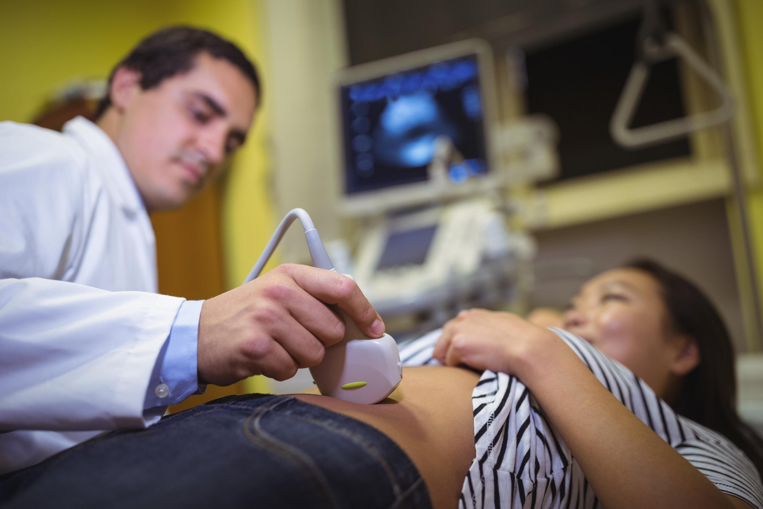
Ultrasound uses high-frequency sound waves that are completely inaudible to the human ear. A handheld device called a transducer or probe emits these sound waves into the body. When the waves hit different tissues, they bounce back at different speeds depending on the density of what they encounter. The machine converts these returning echoes into a real-time image on screen.
Dense structures like bone reflect sound waves strongly and appear bright white on the image. Fluid-filled structures like the bladder or a cyst allow sound waves to pass through easily and appear dark. Solid organs like the liver and kidneys appear in various shades of grey depending on their tissue composition. This is why ultrasound is particularly good at distinguishing between solid masses and fluid-filled ones, a distinction that matters enormously in diagnosis.
No radiation is involved. This makes ultrasound safe for everyone including pregnant women, young children, and patients who need repeated imaging over time. You can find out more about the range of ultrasound scans available at Jhansi Diagnostics & Fetal Medicine Centre on the dedicated page.
What an Abdominal Ultrasound Shows
An abdominal ultrasound is one of the most commonly requested scans. It gives Dr Furquan Ahmad a detailed view of the organs inside the abdomen including the liver, gallbladder, bile ducts, pancreas, spleen, kidneys, and major blood vessels.
In the liver, ultrasound can detect fatty changes, cirrhosis, cysts, haemangiomas, and masses that may require further investigation. In the gallbladder, it is highly accurate at identifying gallstones, even very small ones, as well as thickening of the gallbladder wall and the presence of polyps. In the kidneys, it shows size, shape, the collecting system, stones, cysts, and signs of obstruction. The pancreas, which can be harder to visualise due to overlying bowel gas, is assessed for size, texture, and any visible abnormalities.
An abdominal ultrasound is also used to detect free fluid in the abdomen, which can indicate internal bleeding, infection, or organ rupture in emergency situations. For patients with abdominal pain, bloating, or unexplained symptoms, this scan is usually the first imaging investigation ordered.
What a Pelvic Ultrasound Shows
A pelvic ultrasound is used to examine the organs in the lower abdomen. In women, this includes the uterus, ovaries, fallopian tubes, and the endometrium, which is the lining of the uterus. In men, it can be used to assess the bladder and prostate.
For women, a pelvic ultrasound detects fibroids, ovarian cysts, polycystic ovarian morphology, endometrial thickening, and structural abnormalities of the uterus. It is also used to investigate irregular periods, pelvic pain, difficulty conceiving, and abnormal bleeding. A transvaginal ultrasound, where the probe is gently placed inside the vaginal canal, gives a clearer and more detailed view of the uterus and ovaries than a transabdominal scan and is often used alongside or instead of it for better accuracy.
What a Pregnancy Ultrasound Shows
Pregnancy ultrasounds are among the most detailed and emotionally significant scans performed at Jhansi Diagnostics & Fetal Medicine Centre. At different stages of pregnancy, the scan shows different things.
In the first trimester, an early scan confirms the pregnancy is inside the uterus, establishes the gestational age, checks for a heartbeat, and identifies whether the patient is carrying more than one baby. The NT scan at 11 to 14 weeks adds a measurement of the fluid at the back of the baby’s neck as part of chromosomal risk screening.
In the second trimester, the anomaly scan at 18 to 20 weeks examines the baby’s entire anatomy in detail. Every major organ system is assessed, the placental position is checked, the amniotic fluid level is measured, and the baby’s growth is compared against standard charts for that gestational age.
In the third trimester, growth scans track the baby’s weight, head circumference, abdominal circumference, and femur length to ensure development is on track. Doppler studies assess blood flow through the placenta and umbilical cord. More information about fetal medicine scanning is available on the fetal medicine page.
What a Doppler Ultrasound Shows
A Doppler ultrasound is a specialised type of scan that measures blood flow rather than just structural images. It uses the Doppler effect, the change in frequency of sound waves as they bounce off moving objects, in this case red blood cells, to calculate the speed and direction of blood moving through vessels.
Colour Doppler adds colour coding to the image to show the direction of blood flow at a glance. Red typically indicates flow towards the probe and blue indicates flow away from it. This helps Dr Furquan Ahmad identify blockages, abnormal flow patterns, varicose veins, deep vein thrombosis, and in pregnancy, compromised blood flow through the placenta and umbilical cord. A Doppler study is often performed as an addition to a standard ultrasound rather than as a standalone scan.
What a Musculoskeletal Ultrasound Shows
Musculoskeletal ultrasound examines the soft tissues around joints including tendons, ligaments, muscles, and bursae. It is particularly useful for diagnosing rotator cuff tears in the shoulder, tennis elbow, plantar fasciitis in the heel, and tendon injuries in the knee and ankle.
Unlike an MRI, a musculoskeletal ultrasound can be performed dynamically, meaning Dr Furquan Ahmad can ask the patient to move the joint while imaging it, which helps identify problems that only appear during movement. It is also faster and more accessible than MRI for many patients. For anyone with joint pain, a sports injury, or unexplained soft tissue swelling, a musculoskeletal ultrasound is often the most practical starting point.
What a Thyroid Ultrasound Shows
A thyroid ultrasound examines the thyroid gland in the neck for size, shape, texture, and the presence of nodules. Thyroid nodules are extremely common and the vast majority are benign, but ultrasound helps characterise their features, size, and vascularity to determine whether further investigation such as a fine needle aspiration is needed.
Thyroid ultrasound is also used to monitor known nodules over time, assess for enlargement of the gland, and investigate symptoms like a visible lump in the neck, difficulty swallowing, or hoarseness of voice.
What Ultrasound Cannot Show
Being clear about the limitations of ultrasound is as important as understanding what it shows. Ultrasound does not image bone well, which is why fractures require an X-ray. It is limited in its ability to assess structures obscured by gas or bone, which is why the pancreas can sometimes be difficult to fully visualise. It does not show the fine detail of brain or spinal cord tissue, for which an MRI is more appropriate.
When Dr Furquan Ahmad cannot fully assess a structure due to technical limitations, this is clearly documented in the report and an alternative imaging approach is recommended. This transparency is an important part of what makes a report at Jhansi Diagnostics & Fetal Medicine Centre clinically useful rather than misleadingly reassuring. If you have questions about which scan is right for your symptoms, the FAQs page is a helpful starting point, or you can explore the full range of our services available at the centre.
Book Your Ultrasound in Jhansi
Whether you have been referred by your doctor or want to book a scan proactively, Jhansi Diagnostics & Fetal Medicine Centre offers the full range of ultrasound scans performed and interpreted personally by Dr Furquan Ahmad. Results are available the same day and findings are explained clearly before you leave.
Book your appointment through the website or get in touch through the contact us page with any questions.