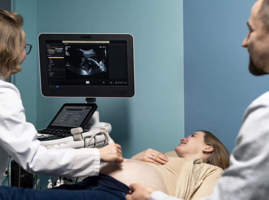
Congenital heart defects are the most common type of birth defect worldwide. They affect the structure of the baby’s heart and can range from small holes that close on their own to complex abnormalities that require surgery shortly after birth. What many families don’t know is that a significant number of these conditions can be identified before birth, during pregnancy, through careful and detailed fetal heart assessment. At Jhansi Diagnostics & Fetal Medicine Centre, Dr Furquan Ahmad, MBBS MD, performs fetal cardiac assessment as part of every detailed anomaly scan, giving families in Jhansi and across Bundelkhand the earliest possible warning of any structural heart concern so that the right preparations can be made before delivery.
Here is how fetal medicine specialists detect heart defects before birth and why early diagnosis matters so profoundly.
Why the Fetal Heart Is Difficult to Assess
The fetal heart is a remarkable structure. By 6 weeks of pregnancy it is already beating. By 20 weeks, when the anomaly scan is performed, it is fully formed and beating at approximately 120 to 160 times per minute. But assessing it on ultrasound is genuinely challenging. It is small, it moves continuously with every heartbeat, and the baby itself is also moving. The structures inside it, the four chambers, the valves, the great vessels emerging from it, are tiny and require both excellent equipment and significant experience to visualise and interpret correctly.
This is one of the reasons fetal cardiac assessment is one of the most demanding parts of the anomaly scan. A quick glance at the heart to confirm it has four chambers is not sufficient to rule out a significant cardiac defect. Many of the most clinically important heart abnormalities, including transposition of the great arteries, coarctation of the aorta, and certain septal defects, can only be detected when the outflow tracts and great vessels are systematically examined alongside the four-chamber view. Dr Furquan Ahmad performs this extended cardiac assessment as a standard part of every anomaly scan at Jhansi Diagnostics & Fetal Medicine Centre. You can read more about the centre’s anomaly scanning services on the fetal medicine page.
What a Normal Fetal Heart Assessment Covers
A thorough fetal cardiac assessment on ultrasound covers several specific views of the heart, each revealing different structures and ruling out different types of abnormality.
The four-chamber view is the starting point. It shows the heart from a cross-sectional angle and allows assessment of the size of the four chambers, the thickness of the walls between them, the position of the heart within the chest, and the opening and closing of the mitral and tricuspid valves. A normal four-chamber view rules out several types of serious cardiac abnormality but not all of them.
The left ventricular outflow tract view shows the aorta arising from the left ventricle. The right ventricular outflow tract view shows the pulmonary artery arising from the right ventricle. These views are critical because some of the most serious congenital heart defects involve abnormalities in how these great vessels connect to the heart, and these abnormalities are invisible on the four-chamber view alone.
The three-vessel view and three-vessel trachea view assess the relative sizes and arrangement of the main vessels above the heart. Abnormalities in these views can indicate coarctation of the aorta, interrupted aortic arch, and other great vessel abnormalities that require neonatal cardiac surgery.
Colour Doppler is used throughout the cardiac assessment to visualise blood flow through the heart and detect abnormal flow patterns that indicate valve problems, septal defects, or outflow obstruction. The combination of two-dimensional imaging and Colour Doppler gives Dr Furquan Ahmad the most complete picture of fetal cardiac anatomy and function available through ultrasound. You can find out more about Colour Doppler services on the ultrasound page.
What Heart Defects Can Be Detected Before Birth?
The range of congenital heart defects detectable on prenatal ultrasound has expanded significantly with improvements in imaging technology and specialist training. While no prenatal ultrasound can guarantee the detection of every possible cardiac abnormality, the following conditions are among those that an experienced fetal medicine specialist can identify before birth.
Ventricular septal defects, which are holes between the two lower chambers of the heart, are the most common congenital heart defect. Large ones are detectable on ultrasound. Very small ones may not be visible prenatally but often close spontaneously after birth without intervention.
Hypoplastic left heart syndrome is a serious condition where the left side of the heart is severely underdeveloped. It is one of the most reliably detected cardiac defects on prenatal ultrasound because the asymmetry between the two sides of the heart is clearly visible on the four-chamber view.
Transposition of the great arteries, where the aorta and pulmonary artery are connected to the wrong ventricles, is detectable on the outflow tract views. This condition requires surgery in the first days of life, and knowing about it before birth allows delivery to be planned at a centre with neonatal cardiac surgery facilities.
Tetralogy of Fallot, a combination of four cardiac abnormalities including a ventricular septal defect and narrowing of the pulmonary outflow tract, is detectable on detailed cardiac assessment. Pulmonary atresia, atrioventricular septal defects, and coarctation of the aorta are among the other conditions that an experienced specialist can identify prenatally.
What Increases the Risk of a Fetal Heart Defect?
While congenital heart defects can occur in any pregnancy, certain factors increase the likelihood and should prompt a more detailed fetal cardiac assessment than the standard anomaly scan.
A family history of congenital heart disease, particularly in a first-degree relative or a previous child, increases the risk in subsequent pregnancies. Chromosomal conditions such as Down syndrome, Edwards syndrome, and Patau syndrome are associated with a high rate of cardiac abnormalities. An increased nuchal translucency measurement on the NT scan is associated with a higher risk of cardiac defects even when chromosomal analysis is normal.
Maternal conditions including pre-gestational diabetes, phenylketonuria, and autoimmune conditions such as systemic lupus erythematosus are associated with specific types of fetal heart abnormality. Exposure to certain medications or infections in the first trimester, when the heart is forming, can also increase the risk.
If any of these risk factors apply to your pregnancy, Dr Furquan Ahmad at Jhansi Diagnostics & Fetal Medicine Centre will perform an extended fetal cardiac assessment and may recommend a dedicated fetal echocardiogram, a more detailed and time-intensive cardiac ultrasound specifically focused on the heart, at a specialist centre. The FAQs page has more information on what these assessments involve.
What Happens If a Heart Defect Is Suspected?
If the anomaly scan raises a concern about the fetal heart, the first step is always to confirm the finding with a more detailed assessment before communicating a diagnosis to the family. Dr Furquan Ahmad examines the heart from multiple angles, applies Colour Doppler, and reviews all available views carefully before drawing any conclusions.
If a cardiac abnormality is confirmed or strongly suspected, the family is informed clearly and compassionately. The nature of the finding, what it means structurally, what the likely postnatal implications are, and what further investigations are recommended are all explained in plain language. A referral for a dedicated fetal echocardiogram at a specialist paediatric cardiac centre is typically the next step for any significant cardiac finding.
Knowing about a heart defect before birth allows families and medical teams to prepare appropriately. Delivery can be planned at a centre with the appropriate neonatal cardiac facilities. The neonatal team can be briefed in advance so that the right support is available from the moment the baby is born. In some conditions, knowing the diagnosis prenatally allows medications to be given to the mother that benefit the baby’s cardiac function before birth. The full range of diagnostic and monitoring services available at Jhansi Diagnostics & Fetal Medicine Centre is outlined on the our services page.
Why Early Detection Changes Everything
For families whose baby is diagnosed with a congenital heart defect before birth, the prenatal diagnosis is not the worst outcome. It is the best possible starting point for managing it. A baby born with an undiagnosed complex heart defect in a hospital without cardiac facilities, without a prepared neonatal team, and without a surgical plan in place is in a far more dangerous position than a baby whose condition was identified at 20 weeks and whose delivery was carefully planned months in advance.
Dr Furquan Ahmad has seen the difference that early cardiac diagnosis makes to outcomes for families in Jhansi and across Bundelkhand. It is one of the primary reasons he performs extended fetal cardiac assessment as a standard part of every anomaly scan rather than limiting the cardiac view to the minimum required by basic protocols.
If you are booked for an anomaly scan or have been referred for a detailed fetal cardiac assessment, you can book your appointment at Jhansi Diagnostics & Fetal Medicine Centre through the website. For any questions before your visit, the team is available through the contact us page. You can also read more about Dr Furquan Ahmad and the centre’s approach on the about us page.
A heart defect found before birth is a heart defect that can be prepared for. That preparation saves lives.