When a baby is growing inside the womb but not gaining weight or size at the expected rate, doctors call it intrauterine growth restriction, commonly referred to as IUGR. It is one of the most serious complications that can affect a pregnancy, and it is also one of the most important to detect early. A baby affected by IUGR is not simply small. It is a baby whose growth has been restricted by an underlying problem, most commonly an issue with the placenta that is preventing adequate oxygen and nutrients from reaching the baby. At Jhansi Diagnostics & Fetal Medicine Centre, Dr Furquan Ahmad, MBBS MD, diagnoses and monitors IUGR pregnancies with the level of specialist attention this condition demands, giving families in Jhansi and across Bundelkhand access to expert fetal care close to home.
Here is everything you need to understand about IUGR, how it is detected, and how it is managed.
What Is Intrauterine Growth Restriction?
Intrauterine growth restriction is a condition where a baby fails to reach its genetically determined growth potential inside the womb. It is typically defined as a baby whose estimated weight falls below the 10th percentile for its gestational age, meaning it is smaller than 90 percent of babies at the same stage of pregnancy. However, not every small baby has IUGR. Some babies are constitutionally small, meaning they are naturally petite but healthy and growing normally on their own growth curve. The distinction between a constitutionally small baby and one with true IUGR is critical, and it is one that requires careful assessment by an experienced fetal medicine specialist.
True IUGR involves a baby that is not just small but is being deprived of the resources it needs to grow. This deprivation has consequences beyond size. A baby with IUGR is at higher risk of stillbirth, preterm birth, low oxygen levels during labour, and a range of health problems in the newborn period and beyond. Early identification and close monitoring are the cornerstones of managing this condition safely.
What Causes IUGR?
The most common cause of IUGR is placental insufficiency. The placenta is responsible for transferring oxygen and nutrients from the mother’s bloodstream to the baby. When the placenta does not develop normally, implants in a suboptimal location, or is affected by disease, its ability to supply the baby adequately is compromised. The baby, receiving less than it needs, slows its growth in response.
Maternal conditions that affect blood flow and vascular health are strongly associated with IUGR. Pre-eclampsia, chronic hypertension, diabetes, kidney disease, autoimmune conditions, and severe anaemia can all impair placental function and restrict fetal growth. Infections during pregnancy, including certain viral infections, can also affect fetal growth directly.
Multiple pregnancies carry a higher risk of IUGR, particularly in identical twins sharing a placenta where one twin may receive a disproportionately smaller share of the placental blood supply. Lifestyle factors including smoking, alcohol use, and poor nutritional intake during pregnancy are also associated with increased risk. In some cases, IUGR is caused by a chromosomal or structural abnormality in the baby itself rather than a placental problem.
How Is IUGR Detected?
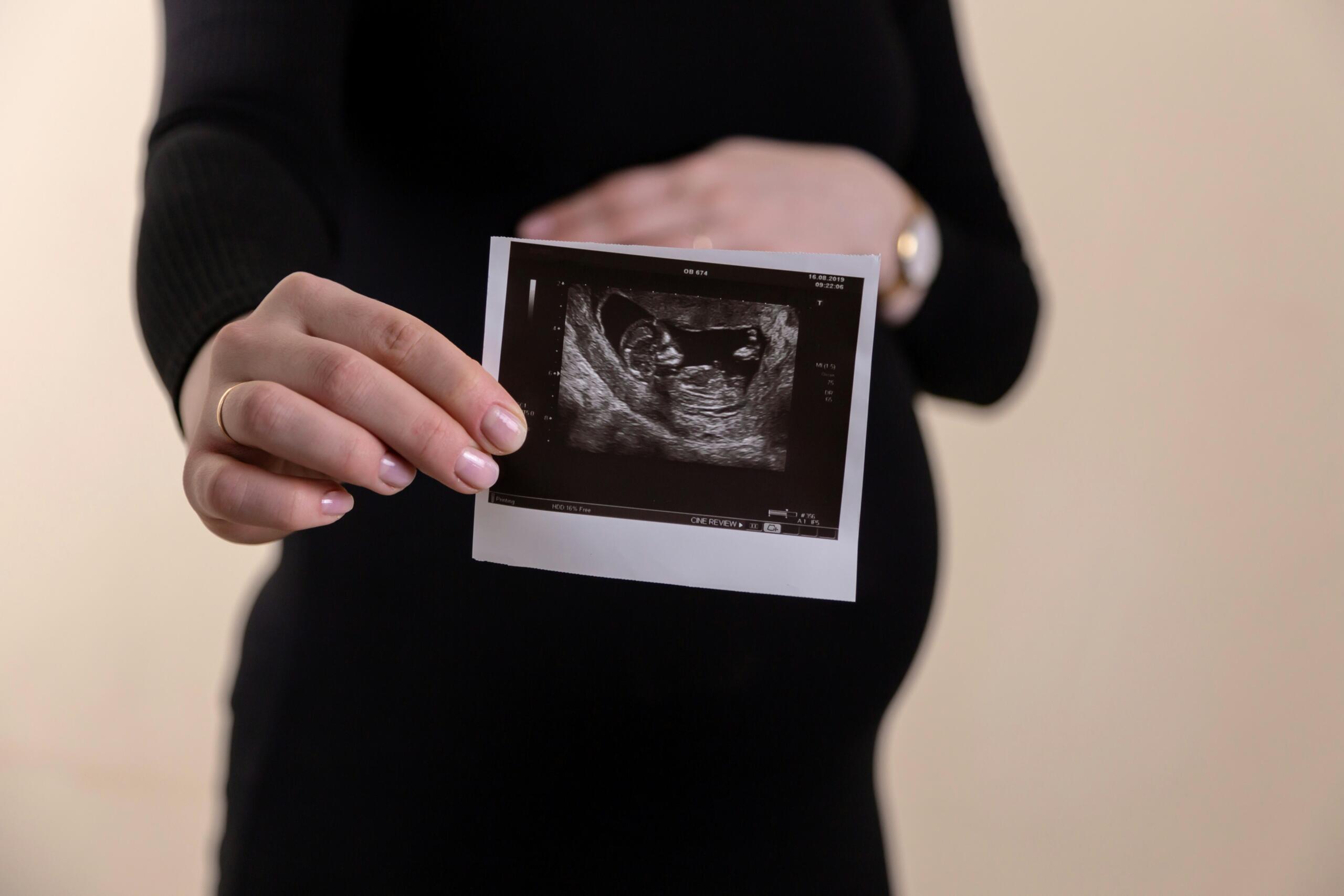
IUGR is detected through careful ultrasound monitoring during pregnancy. Dr Furquan Ahmad uses a combination of fetal biometry, Doppler blood flow studies, and amniotic fluid assessment to diagnose and monitor IUGR at Jhansi Diagnostics & Fetal Medicine Centre.
Fetal biometry involves measuring the baby’s head circumference, abdominal circumference, femur length, and estimated weight and plotting these measurements against standard growth charts for the gestational age. A baby whose measurements fall below the 10th percentile, or whose growth has slowed significantly between scans even if still within the normal range, raises the possibility of IUGR.
Doppler studies are the most important tool in distinguishing true IUGR from constitutional smallness and in assessing the severity of the condition. The umbilical artery Doppler measures blood flow resistance in the cord. As placental insufficiency worsens, resistance increases and the blood flow pattern changes in characteristic ways. Reduced end-diastolic flow is an early sign. Absent end-diastolic flow is a more serious finding indicating significant placental compromise. Reversed end-diastolic flow is a critical finding that requires immediate obstetric review and often delivery.
The middle cerebral artery Doppler adds further information about how the baby is coping. When a baby with IUGR is under stress, it redirects blood flow to protect its brain at the expense of other organs. This brain-sparing response is visible on Doppler and is an important marker of fetal wellbeing. The ductus venosus Doppler, which assesses blood flow through a vessel near the baby’s heart, is used in more advanced cases to assess cardiac function and help time delivery decisions.
Amniotic fluid volume is also assessed at every monitoring scan. Reduced amniotic fluid, known as oligohydramnios, is a common accompaniment to IUGR and reflects reduced fetal urine output caused by decreased blood flow to the kidneys. You can read more about the ultrasound and fetal medicine services available at the centre on the dedicated pages.
Who Is at Risk and When Should Monitoring Begin?
Certain groups of pregnant women are at higher risk of IUGR and should begin fetal growth monitoring earlier and more frequently than the standard antenatal schedule. Women with pre-eclampsia or chronic hypertension, women with a previous pregnancy affected by IUGR or stillbirth, women carrying twins sharing a placenta, women with diabetes or autoimmune conditions, and women who smoke during pregnancy are all in this higher-risk category.
Dr Furquan Ahmad recommends that women in these groups have a uterine artery Doppler assessment at 20 to 24 weeks, which can identify early signs of abnormal placental blood flow before growth restriction becomes apparent on biometry. This early warning allows monitoring to be intensified at the right time rather than playing catch-up after growth restriction has already set in.
For women without specific risk factors, growth monitoring scans are typically introduced in the third trimester if clinical examination suggests the baby may be small. Any woman whose fundal height measurement is lagging behind her gestational age should be referred for a growth scan promptly rather than waiting for the next routine appointment.
How Is IUGR Managed?
There is no treatment that can directly reverse IUGR or restore placental function. Management is therefore focused on close monitoring to detect any deterioration in fetal wellbeing and timing delivery to balance the risks of prematurity against the risks of continuing the pregnancy with a compromised placenta.
The frequency of monitoring depends on the severity of the IUGR and the Doppler findings. Mild IUGR with normal Doppler studies may require growth scans every two weeks. More significant IUGR with abnormal umbilical artery Doppler findings may require twice-weekly or even daily monitoring in a hospital setting as the pregnancy approaches term.
Dr Furquan Ahmad works closely with the referring obstetricians of his patients at Jhansi Diagnostics & Fetal Medicine Centre, providing detailed scan reports and Doppler assessments that guide the obstetric team’s decisions about when to deliver. The goal is always to keep the baby in the womb as long as it is safer than being born, and to deliver before the placental compromise puts the baby at serious risk.
Delivery planning in IUGR pregnancies takes into account gestational age, the severity of Doppler abnormalities, amniotic fluid levels, biophysical profile scores, and the availability of neonatal care for a preterm baby. These decisions are complex and require experienced specialist input at every stage.
Mothers with IUGR pregnancies are also advised on lifestyle measures that support the best possible outcome: adequate rest, avoidance of smoking and alcohol, good nutrition, and prompt reporting of any reduction in fetal movements. Reduced fetal movements are a warning sign that should never be ignored in an IUGR pregnancy and always warrant same-day assessment.
What Happens After Delivery?
Babies born after an IUGR pregnancy may need additional support in the neonatal period depending on their gestational age and birth weight. They are at higher risk of low blood sugar, temperature instability, breathing difficulties, and infection in the immediate newborn period. With appropriate neonatal care, many IUGR babies go on to catch up in growth and development during infancy and early childhood.
Long-term follow-up is important for children born with IUGR, as research has linked severe early-onset growth restriction with a higher risk of metabolic and cardiovascular conditions in adult life. Awareness of this risk allows parents and paediatricians to monitor growth and development proactively from the start.
For families in Jhansi who have received an IUGR diagnosis or who are concerned about their baby’s growth, the FAQs page at Jhansi Diagnostics & Fetal Medicine Centre addresses common questions. The full range of monitoring services is outlined on the our services page. Appointments can be booked through the booking appointment page, and the team is available through the contact us page for any questions before your visit.
IUGR is a condition that demands expert attention and consistent monitoring. With the right specialist involvement from the right point in the pregnancy, the outcomes for mother and baby are significantly better than when it is managed without specialist input.