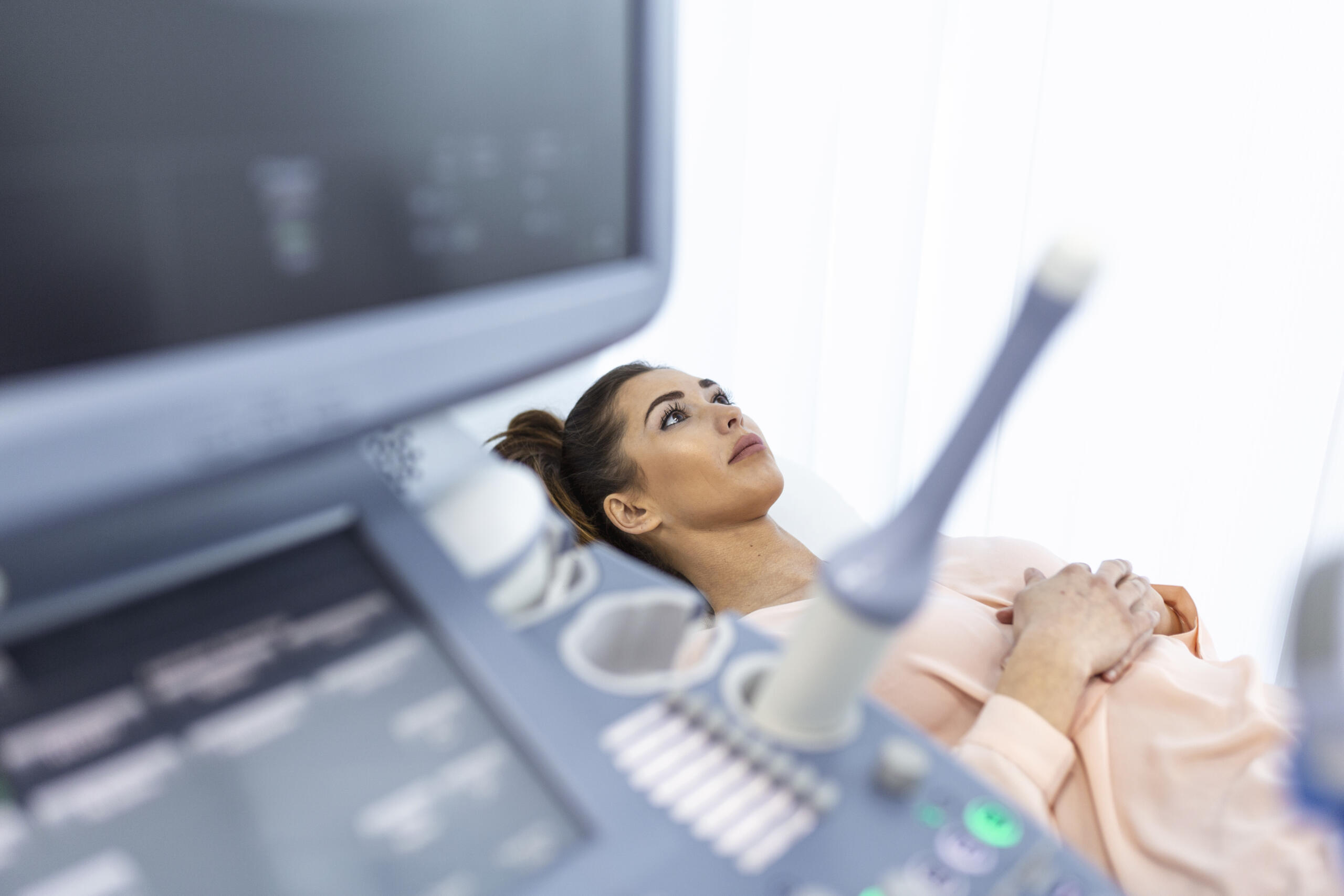
Ultrasound
Our Services
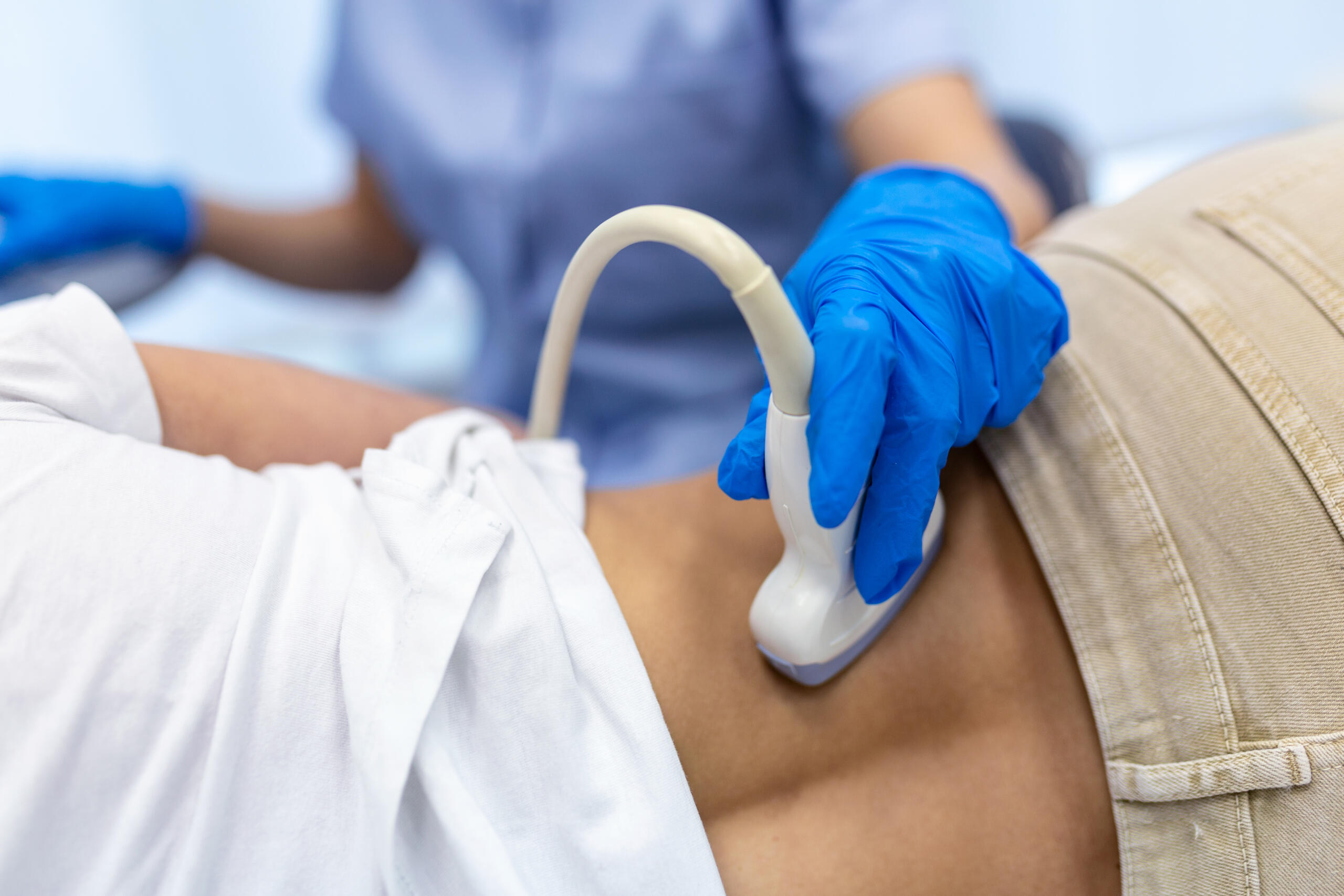
Safe & Accurate Ultrasound Diagnostics
At Jhansi Diagnostics & Fetal Medicine, we offer advanced ultrasound services using state-of-the-art technology to provide accurate, real-time imaging for diagnosis and monitoring
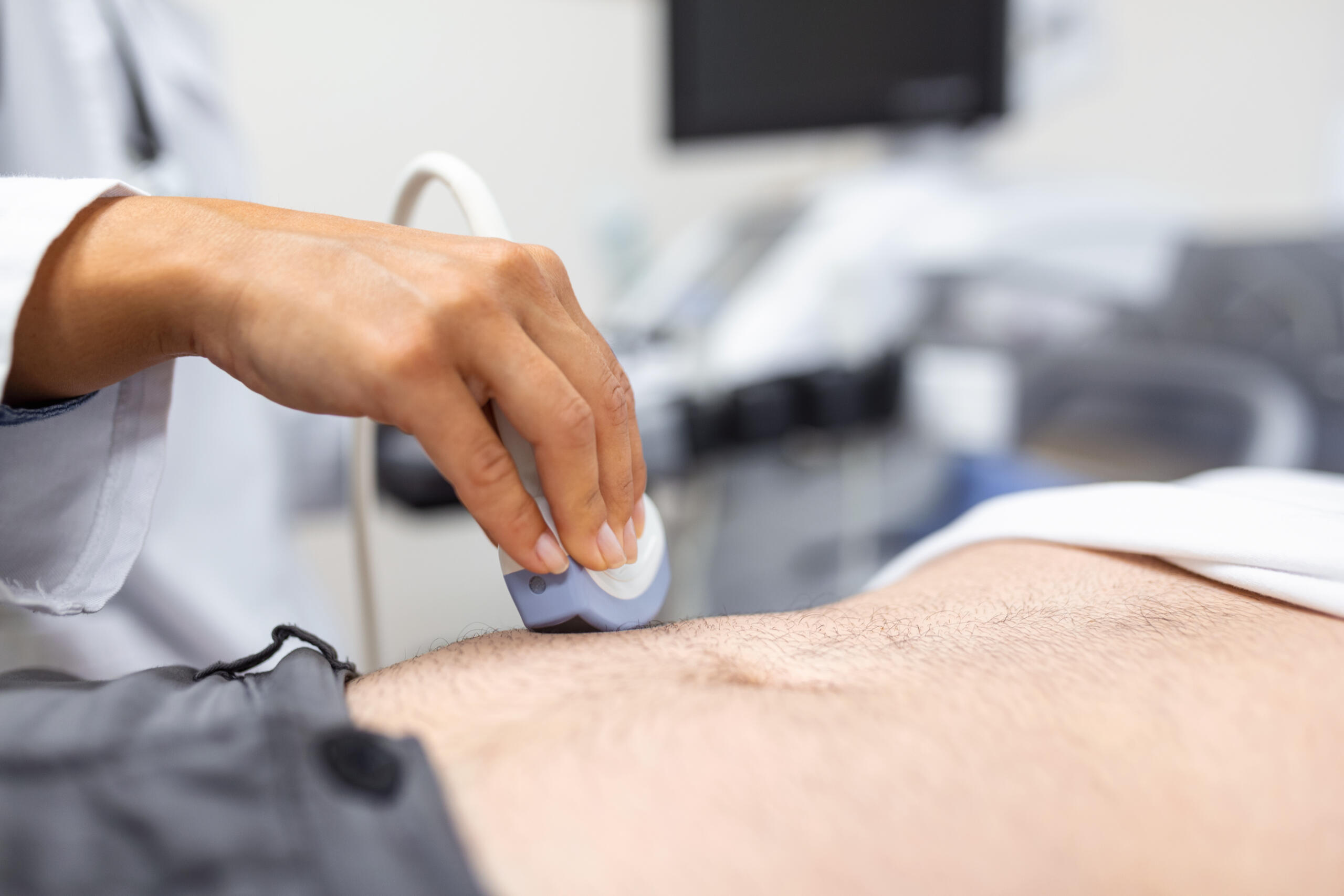
Abdominal Ultrasound
Liver, kidneys, gallbladder, pancreas, and spleen assessment.
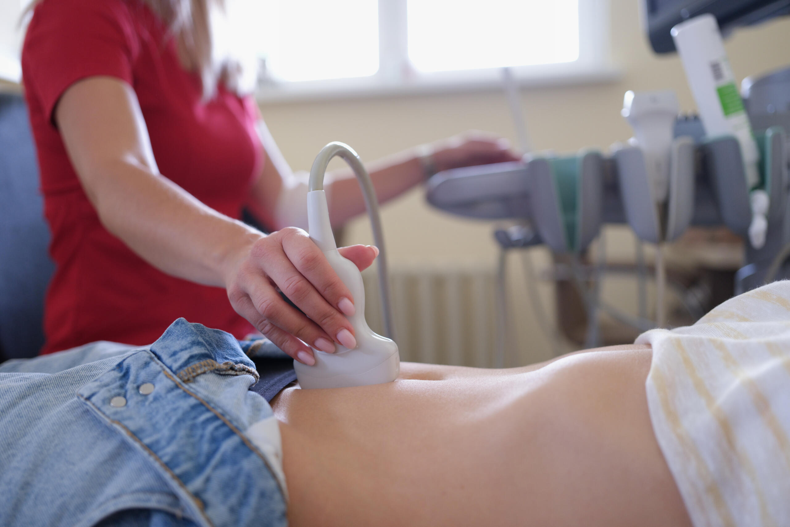
Pelvic Ultrasound
Reproductive organs, bladder, and gynecological evaluation.

Pregnancy Ultrasound
Fetal monitoring and development with zero radiation.
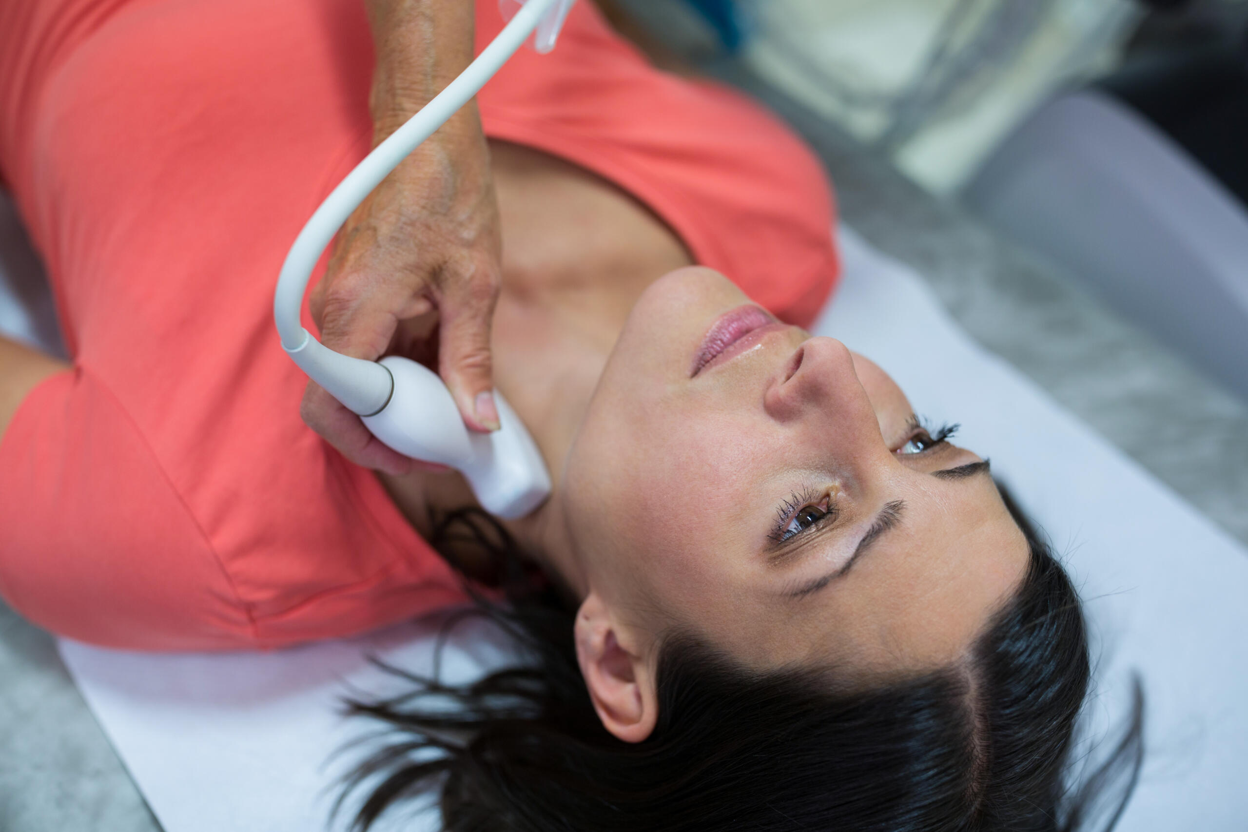
Thyroid Ultrasound
Nodules, cysts, and gland assessment.
Breast Ultrasound
Lump evaluation and tissue assessment without radiation.
Doppler Ultrasound
Blood flow and vascular assessment in real time.
What is an Ultrasound Scan?
An ultrasound scan, also known as sonography, is a widely used imaging technique that employs high-frequency sound waves to create images of structures inside the body. It is a non-invasive, painless, and radiation-free method for diagnosing, monitoring, and treating various medical conditions. From prenatal care to detecting abnormalities in internal organs, the ultrasound scan plays a crucial role in modern medicine.
This article provides an in-depth understanding of ultrasound scans, including their purpose, how they work, test results interpretation, normal ranges, preparation tips, and answers to common patient questions.
An ultrasound scan is a diagnostic imaging test that uses sound waves to produce real-time images of tissues, organs, and blood flow within the body.
How It Works:
- A device called a transducer emits sound waves that bounce off internal tissues.
- The reflected sound waves are captured and converted into images by a computer.
Types of Ultrasound Scans:
- External Ultrasound: Performed by moving the transducer over the skin.
- Internal Ultrasound: Involves inserting a specialized transducer into the body (e.g., transvaginal or transrectal ultrasound).
- Doppler Ultrasound: Measures blood flow through vessels.
Why is an Ultrasound Scan Important?
Ultrasound scans are essential diagnostic tools due to their safety, versatility, and effectiveness
Non-Invasive and Radiation-Free
Provides detailed imaging without exposing the patient to radiation.
Real-Time Results
Offers live imaging for procedures like biopsies or monitoring pregnancies
Wide Applications
Used for a variety of medical conditions, from prenatal care to detecting tumors or gallstones.
Accessible and Affordable
Widely available in clinics, hospitals, and diagnostic centers.
Precision Ultrasound for Accurate Diagnosis
At Jhansi Diagnostics & Fetal Medicine, we provide high-quality ultrasound imaging using advanced technology to deliver accurate diagnoses and support better healthcare decisions. Our expert radiologists ensure every scan is performed with precision, care, and patient comfort in mind.
Ultrasound is a safe, non-invasive imaging technique that uses sound waves to create detailed images of internal organs, tissues, and fetal development. With state-of-the-art equipment and timely reporting, we are committed to delivering reliable diagnostic services for patients of all ages.
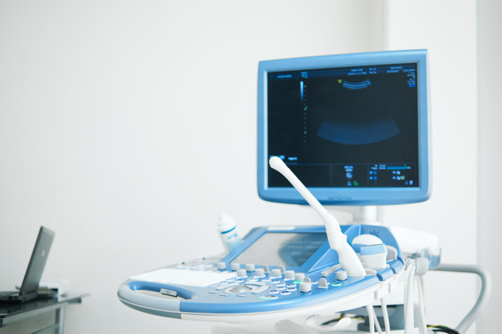
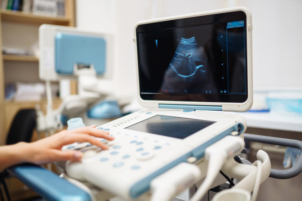
Jhansi Diagnostics & Fetal Medicine is one of the most advanced imaging centres in the Bundelkhand region, equipped with the first GE Voluson Signature 18 Expert Ultrasound System in Bundelkhand and adjoining areas. This state-of-the-art technology delivers exceptional image clarity, advanced fetal assessment, and highly accurate diagnostic results, making us a trusted destination for advanced ultrasound and fetal medicine services.
- Zero Radiation
- Fast Results
- Real-Time Imaging
Features
Why Choose Our Ultrasound Services?
At Jhansi Diagnostics & Fetal Medicine, we combine advanced ultrasound technology with expert clinical expertise to deliver accurate, safe, and reliable diagnostic imaging. Our commitment to quality care ensures precise results and a comfortable experience for every patient.
Fast Reporting
Timely and reliable reports to support quick medical decisions.
Patient-Centered Care
Comfortable, compassionate, and personalized diagnostic experience.
Real-Time Imaging
Live visualization for better diagnostic assessment and monitoring.
Zero Radiation
Safe imaging using sound waves without harmful radiation exposure.
Abdominal Ultrasound
Fast for 6–8 hours before the scan and avoid food or carbonated beverages. Small sips of water may be allowed if advised by your healthcare provider.
Pelvic Ultrasound
Drink 4–6 glasses of water one hour before your appointment and avoid emptying your bladder until the scan is completed. A full bladder helps improve image clarity and ensures more accurate diagnostic results.
Internal Ultrasound Examinations
Follow the preparation instructions provided by your healthcare provider. Additional preparation may be required depending on the type of internal ultrasound being performed.
Patient Preparation
Patient Preparation for Different Ultrasound Scans
To ensure accurate and high-quality imaging, certain ultrasound examinations may require specific preparation before your appointment. Please follow the guidelines below based on the type of scan prescribed.
Pregnancy Ultrasound
Follow the instructions provided during appointment scheduling. Some early pregnancy scans may require a full bladder to ensure clear imaging and accurate assessment of fetal development.
Doppler Ultrasound
Wear comfortable, loose-fitting clothing and follow any specific preparation instructions provided by your doctor before the examination.
Cardiac Ultrasound (Echocardiography)
No special preparation is usually required for a cardiac ultrasound. You can continue taking your regular medications unless your doctor advises otherwise.
General Preparation Guidelines
Carry your doctor's prescription and any previous medical reports, wear comfortable clothing for easy examination, avoid applying lotions or creams to the area being scanned, and arrive at least 15 minutes before your scheduled appointment.
Who Should Get Ultrasound?
Ultrasound is ideal when your doctor needs to see soft tissues, organs, or blood flow without radiation. It’s particularly valuable for pregnancy, frequent monitoring, and when other imaging methods aren’t suitable.
Pregnant women for fetal monitoring and development
Abdominal pain, liver, kidney, or gallbladder concerns
Pelvic pain or gynecological symptoms
Breast lumps or tissue concerns
Thyroid nodules, enlargement, or suspected thyroid disease
Blood flow or vascular assessment
Tendon injuries or soft tissue evaluation
Testicular pain, lumps, or swelling requiring scrotal evaluation
Guided procedures such as biopsies, aspirations, or injections
Infant hip screening for developmental dysplasia in newborns
Nuchal Translucency (NT) Scan
The Nuchal Translucency (NT) Scan is performed between 11 and 13 weeks 6 days of pregnancy to assess early fetal development and screen for chromosomal conditions, including Down syndrome and major fetal abnormalities.
Why is an NT Scan Important?
Accurate Pregnancy Dating : Helps determine the baby’s gestational age and estimated delivery date.
Detection of Multiple Pregnancies : Identifies twins or multiple pregnancies and assesses placental sharing.
Early Fetal Assessment : Screens for major structural abnormalities visible during early pregnancy.
Assessment of Chromosomal Risk : Evaluates the risk of Down syndrome and other chromosomal conditions.
Advanced Risk Evaluation : Combines ultrasound findings with maternal age and blood test results for comprehensive screening.
Assessment of Chromosomal Risk : Evaluates the risk of Down syndrome and other chromosomal conditions.
FAQs About Ultrasound Scans
Ultrasound scans are used to diagnose, monitor, and guide treatment for a wide range of conditions. They are commonly used in pregnancy, abdominal imaging, cardiac assessment, and detecting soft tissue injuries or blood flow issues.
No, ultrasound scans are painless. You may feel slight pressure when the transducer is moved over the skin or inserted for internal scans, but the procedure is generally comfortable.
Most scans are completed within 15–30 minutes. Some complex procedures, like Doppler ultrasounds, may take slightly longer.
Fasting is required for certain types of ultrasounds, such as abdominal scans, to reduce gas and improve image clarity. Your doctor will provide specific instructions.
Yes, ultrasounds are completely safe during pregnancy as they do not use radiation. They are the primary method for monitoring fetal health and development.
It depends on the type of scan. For abdominal ultrasounds, fasting is usually required. For other scans, you can eat and drink normally.
Typically within 30 min - 1 hour.
Wear loose-fitting clothing that can be easily adjusted or removed to access the area being examined.
Yes, you can return to your usual activities immediately unless instructed otherwise by your doctor.